Conference Schedule

Day1: July 23, 2018
Keynote Forum

09:30-10:10
Weisi Yan
University of South Alabama
Title: The application of lattice therapy in clinical radiation oncology
09:30-10:10
Biography
Weisi Yan has finished his PhD in Pharmacology in Weill Graduate School of Cornell Medical College. He was trained in NYP/Cornell for Clinical Radiation Oncology. He subsequently held Faculty appointments at New York-Presbyterian Hospital and New York Hospital Queens. He also served as an Assistant Professor of Clinical Radiology at the Weill Cornell Medical College of Cornell University. His specialties are head and neck cancers, lung cancer, lymphoma and GI cancers. In addition, he has an extensive training in Stereotactic Radiation Therapy, in which high-dose radiotherapy beams are aimed at a tumour from many different directions with a high level of precision. He is also the Director of the Cyber Knife Robotic Radiotherapy System at Mitchell cancer Institute at the University of South Alabama.
Abstract
High-dose GRID radiotherapy, sometimes termed spatially-fractionated GRID radiotherapy (SFGRT), is a treatment modality designed for the treatment of advanced bulky tumours. GRID therapy would induce a more rapid rate of tumour cell apoptosis in bulky, hypoxic, tumours than conventional dosimetric approaches. Indeed, dramatic clinical responses have been reported with GRID radiotherapy. GRID's application is limited by tumour location (only for superficial tumours). Lattice therapy (LRT) is a 3D version of GRID which can create high dose islands within the tumour target with similar dose distribution and effects like brachytherapy with external beam radiation. LRT is not limited by tumour size and location due to its design to have a rapid dose fall-off outside these high dose islands, resulting in low-dose valleys within the tumour as well as low radiation dose outside of the target volume. In vivo data has shown that single fraction, high-dose LRT significantly delayed growth of both local and distant tumours. High-dose LRT induces increased secretion of inflammatory cytokines and abscopal effects in distant tumours untreated with LRT. Clinical data in LRT is promising; a patient with voluminous ovarian carcinosarcoma (size>6 cm) was treated with lattice with excellent local control and no toxicity. LRT combined with chemotherapy for the treatment of the large recurrent ovarian mass in this patient was very well tolerated. In a small group of cancer patients treated with IMRT and LRT (18Gy) as a boost, 75% patients achieved a PR; LRT was well tolerated in this group of patients.

10:10- 10:50
Igor Smit
Ijsselland Ziekenhuis
Title: Percutaneous tissue acquisition under radiological guidance in delicate areas for precision medicine
10:10- 10:50
Biography
Precision medicine has gained wide attention in various disciplines such as oncology. Selection of drugs and patients is imperative to cope with increasing complexity and costs of new systemic treatments. Almost all these advances are based on molecular biology of the target lesion. High quality tissues meaning enough, pure and fresh are prerequisite for these expensive and complex medical innovations. Often, the biopsy step is the bottleneck because of concerns for patient discomfort, morbidity/mortality, and poor tissue quality. While surgery is expensive and cumbersome, tru-cut technologies are inappropriate also because of the relatively small size and the heterogeneity of samples. New macrobiopsy technology can provide samples with sufficient quality in a comfortable out-patient setting. Recently, new progress has been made for biopsy in the most delicate areas of the human body. This study collected cases of patients that were seen in six clinical departments on admission for lesions that were inaccessible for other percutaneous tissue acquisition methods in areas such as head and neck, thorax, liver, intra-abdominal, retroperitoneal, and intrapelvic sites. The biopsy was taken by Spirotome of sizes between 14 and 8 G. The procedure was considered successful if the patient had no side effects and the size of the sample was sufficient for all prospected histological and biomolecular tests. The end-point of this non-randomized study was to detect locations where a macrobiopsy could not be taken. For all areas, considered to be delicate for tissue acquisition, at least one successful procedure was possible. In almost all cases, the procedure could be considered successful. Tips and tricks will be presented. With the appropriate tools for imaging and biopsy, high quality tissue samples can be obtained from all body parts, even the most delicate, providing access to precision medicine for every patient.
Abstract
Precision medicine has gained wide attention in various disciplines such as oncology. Selection of drugs and patients is imperative to cope with increasing complexity and costs of new systemic treatments. Almost all these advances are based on molecular biology of the target lesion. High quality tissues meaning enough, pure and fresh are prerequisite for these expensive and complex medical innovations. Often, the biopsy step is the bottleneck because of concerns for patient discomfort, morbidity/mortality, and poor tissue quality. While surgery is expensive and cumbersome, tru-cut technologies are inappropriate also because of the relatively small size and the heterogeneity of samples. New macrobiopsy technology can provide samples with sufficient quality in a comfortable out-patient setting. Recently, new progress has been made for biopsy in the most delicate areas of the human body. This study collected cases of patients that were seen in six clinical departments on admission for lesions that were inaccessible for other percutaneous tissue acquisition methods in areas such as head and neck, thorax, liver, intra-abdominal, retroperitoneal, and intrapelvic sites. The biopsy was taken by Spirotome of sizes between 14 and 8 G. The procedure was considered successful if the patient had no side effects and the size of the sample was sufficient for all prospected histological and biomolecular tests. The end-point of this non-randomized study was to detect locations where a macrobiopsy could not be taken. For all areas, considered to be delicate for tissue acquisition, at least one successful procedure was possible. In almost all cases, the procedure could be considered successful. Tips and tricks will be presented. With the appropriate tools for imaging and biopsy, high quality tissue samples can be obtained from all body parts, even the most delicate, providing access to precision medicine for every patient.

11:10-11:50
Donglu Shi
CEAS-University of Cincinnati, USA
Title: Detection of circulating tumor cells by electrically-charged magnetic nanoprobes
11:10-11:50
Biography
Donglu Shi is currently the Chair of the Materials Science and Engineering Program at the University of Cincinnati. He received his PhD in Engineering from the University of Massachusetts, Amherst. He was the Staff Scientist in the Materials Science Division of Argonne National Laboratory between 1988 and 1995. He has so far published 270 refereed SCI journal publications including Physical Review Letters, Nature, ACS Nano, and Advanced Materials. His main interests include Nanostructure Design, Nano Biomedicine, Nanophotonics, and Magnetism. The most recent works pioneer several novel approaches in developing multifunctional nano carrier systems for early cancer diagnosis and therapy.
Abstract

11:50-12:30
Kenji Ohe
Fukuoka University, Japan
Title: From understanding the molecular mechanism of aberrant ERαpre-mRNA toward the therapeutic application in breast cancer
11:50-12:30
Biography
Kenji Ohe has completed his PhD at Kyushu University, Japan, and started working as a Postdoctoral fellow at Paolo Sassone Corsi’s lab when it was at IGBMC, France, and Akila Mayeda’s lab when it was at Miami, Florida, USA. He is now working on therapeutical tactics on manipulating alternative splicing as an Associate Professor at Faculty of Pharmaceutical Sciences, Fukuoka University, Japan.
Abstract
HMGA1a (formerly termed HMGI) is known as a DNA-binding transcription factor with oncogenic properties. We have reported that it also binds to RNA in a sequence-specific manner. HMGA1a anchors U1 snRNP to the 5’ splice site of presenilin-2 exon 5 to induce its aberrant exon skipping only when the HMGA1a RNA-binding site is adjacent to the 5’ splice site in sporadic Alzheimer’s disease. In order to seek for other target genes of HMGA1a, we were prompted to search for HMGA1a RNA-binding sites in the estrogen receptor alpha (ERα) gene where both have been extensively studied in breast cancer. We performed a sequence homology search for the presenilin-2 (PS2) HMGA1a RNA-binding site and found a specific sequence 33-nt upstream of the 5’ splice site of ERα exon 1. Therefore, we examined; (i) HMGA1abinding to the RNA sequence by RNA-EMSA, (ii) splicing activity in cultured MCF-7 cells that were transfected with HMGA1a-expression and its RNA decoy expression plasmids, (iii) splicing activity in vitro. We found it switched two alternatively spliced isoforms, ERα66 and ERα46. HMGA1a-mediated U1 snRNP anchoring to the adjacent pseudo-5’ splice site was checked by psoralen-mediated UV crosslinking combined with RNA-EMSA. The effect of the decoy oligonucleotides containing the PS2 HMGA1a RNA-binding site in MCF-7 cells was further checked by transplanting its stable transfectant in nude mice showing increased estrogen-dependent growth. However, in tamoxifen-resistant MCF-7 TAMR1 cells, the HMGA1a RNA-binding decoy oligonucleotides improved tamoxifen-responsiveness by inhibiting estrogendependent cell proliferation. We conclude that this HMGA1a RNA-binding decoy oligonucleotides would be implicated in novel therapeutic application to improve tamoxifen effectiveness in breast cancer where tamoxifen lack effect.
Tracks
- Cancer Research & Cancer vaccines | Biomarker and drug targeting | Clinical Radiational Oncology | Cancer Biology | Immuno Oncology studies |Cancer Research & Cancer Vaccines
Location: Meeting Place 2

Donglu Shi
University of Cincinnati, USA
Chair

Weisi Yan
University of South Alabama, USA
Co Chair

13:30-14:00
Maria del Rosario Davalos Gamboa
University of San Simon, Bolivia
Title: Immune system, food and infectious diseases in a focal sample of children and adolescents with cancer in Cochabamba, Bolivia
13:30-14:00
Biography
Maria Del Rosario Dávalos Gamboa has completed her Pharmaceutical Biochemistry degree from the University Of San Simon Bolivia. She has obtained her PhD in the Pontifical University San Francisco Xavier of Chuquisaca. She has more than 15 scientific articles published in prestigious Journals. She was the Director of the Research Institute of the Faculty of Dentistry UMSS, she won a competency exam at the Faculty of Dentistry UMSS in the Chair of Biochemistry and currently, she is Titular professor of Biochemistry in the same faculty.
Abstract
Objective: To determine the influence of the immune system, food and infectious diseases, for the development of cancer in a focal sample of children and adolescents who usually attend the Manuel Ascencio Villarroel Children's Hospital in the Cochabamba region (Bolivia).
Methods: Cross-sectional study conducted in January and February 2016 in children and adolescents who regularly attend the Manuel Ascencio Villarroel child's Hospital, between the ages of 2 months and 16 years (n = 45) of the Cochabamba region (Bolivia). The parents and / or guardians of the participants were surveyed for a descriptive analysis.
Results: They had two or more signs and symptoms (low immunity) by the weakened immune system 86.67%. They were usually affected by, virus and influenza 51.11%, with muscular pain and constant joints 44.44%, watery eyes and nose running in 35.55%, persistent headache in 40%, they got tired a lot and fatigued despite of rest 35.56%, 31.11% were regularly ill, had a delay in recovering from diseases 28.88%, exhibited a fixed pattern of disease 28.88%, fought endlessly with the disease 24.44%. In the type of food those who consumed natural products before their diagnosis of cancer, 41.66% the use of natural carbohydrates, 61.11% natural protein, 61.11% natural fats, 63.88% of natural fruits, 55 % natural fibers, 27.77% drank a lot of water. In the type of inadequate food that they consumed before their diagnosis of cancer, 33.33% cured meats, 75% soft drinks, 83.3% refined sugar, junk food and refined flour. No statistically significant differences were detected in the influence of infectious diseases.
Conclusions: In the study it was determined that for the development of cancer in children and adolescents of the focal sample, both their weakened immune system and their diet influenced them, although their diet was varied with natural products, usually they consumed sodas, refined sugar, white flour and junk food, foods that are related to the origin of cancer.
14:00-14:30
14:00-14:30
Biography
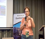
Dina Ron has completed her PhD from Ben-Gurion University and Postdoctoral studies from the National Cancer Institute, NIH, USA. She is a Lab Chief in the Biology Department, the Israel Institute of Technology. She has published more than 60 papers in Refereed professional journals and served as an Editorial Board Member of the Journal of Biological Chemistry.
Abstract
Carcinomas account for over 80% of all human cancer types with no effective therapies. The tumor suppressor Sef [similar expression to fibroblast growth factors (FGF)] is expressed in all the human epithelial tissues and is down-regulated in all the carcinoma types that have been examined so far in a manner that correlates with tumor aggressiveness. We examined the therapeutic potential of restoring the expression of the hSef-b isoform in a prostate carcinoma model. In our in vitro studies, hSef-b inhibited the proliferation of TRAMP C2 cells and attenuated the activation of ERK/MAPK as well as the master transcription factor NF-κB in response to FGF and IL-1/TNF, respectively. Both FGF and NF-κB are strongly implicated in prostate carcinoma progression. Next, the hSef-b, gene was delivered using therapeutic ultrasound (TUS) to pre-established prostate tumors in vivo. Tumors were injected with a bicistronic vector co-expressing hSef-b with eGFP to serve as a reporter for transfection rates, and treated with TUS. Transfection efficiency of plasmid co-expressing hSef-b/eGFP into TRAMP C2 tumors following a single TUS application was 14.7±2.5%. Repeated TUS treatments with hSef-b plasmid, significantly suppressed prostate tumor growth (60%) through inhibition of cell proliferation (60%), and also reduced blood vessel density (56%). In addition, the levels of the promitogenic and proangiogenic factor, FGF2, were significantly reduced following repeated TUS treatments with hSef-b plasmid. Collectively, our results strongly suggest that hSef-b acts in a cell autonomous as well as in a paracrine manner and further revealed the efficacy of non-viral, TUS-based hSef-b gene delivery approach for the treatment of prostate cancer tumors, and possibly other carcinomas where Sef is downregulated. Moreover, using this approach, we discovered that hSef-b is also furnished with the capacity to inhibit tumor angiogenesis.

14:30-15:00
Slobodnikova Jana
Alexander Dubcek University of Trencin, Slovakia
Title: Intervention methods in patients after surgical therapy of breast cancer: the retrospective study
14:30-15:00
Biography
Jana Slobodníková has completed Specialization of Radiology I and II from Charles University in Prague, Faculty of Medicine. She has completed Post-doctorate and CSc (PhD) at the Institute of Experimental Oncology, Slovak Academy of Sciences. She was an Assistant Professor at Tyrnaviensis University, Hosting Professor at St. Elizabeth High School in Bratislava. She has been teaching at several universities in Prague, TrenÄín, Trnava and Bratislava. She is Author of teaching text, university textbooks, and lead workshops. She organizes international congresses and workshops. She has published more than 90 scientific papers, from then 25 papers in renowned journals and has been active as Editorial Board Member of repute in 5 scientific periodicals. She has published 3 monographies, and has founded the Section of Breast Imaging in 1996. Since 1996, she is the President of the Section of Breast Imaging of Slovak radiologic Society and Vice President of the Slovak Society of Ultrasound in Medicine. She has Membership in organizations: ECR, EUSOBI, EFSUMB, SSUM (Slovak Society of Ultrasound in Medicine) and SRS (Slovak Radiology Society).
Abstract
Introduction: In the Slovak Republic, breast cancer is the most common type of oncological disease in women, the leading cause of cancer mortality in all women. In the past 10 years, breast cancer mortality has continued to slowly decrease; the breast cancer incidence in the past 20 years has continued to increase, likely due to program of preventive mammography
The Aim: The aim of this retrospective study was to asses the importance, the position of the interventional methods; core cut biopsy and fine needle aspiration biopsy under ultrasound control in the differential diagnostics of changes after surgical treatment for breast cancer
Patients & Methods: We evaluated examinations of 385 patients with newly diagnosed breast carcinoma after conservative surgical therapy in period between January 2005 and December 2016 from Clinic of Radiology in TrenÄín. In all the cases, we performed ultrasound, mammography 2D and 3D examinations, ultrasound guided biopsy with free hand methods, needle 14, 16 and 20 G.
Results: The major findings as postoperative changes were seromas, haematomas, fat necrosis, skin thickening, inflammatory changes, granulomas, postoperative scars and local tumor recurrence.
Conclusion: The ultrasound guided interventional methods; fine needle aspiration cytology and core cut biopsy are reliable and perfect methods for the differential diagnosis of postoperative changes and the follow up of postoperative changes.

15:00-15:30
Biography
George Kunudji has attained his PhD from the University of Ghana, Legon and Postdoctoral studies from University of Ghana Medical School. He is the Director of Bikbok Herbal Centre, a reputable herbal organisation that is dispensing services across the country, Ghana and currently discovered herbal antidote to the treatment of cancer tumour. Moreover, he has been serving as the Chairman of the health advocacy group of Asuboa Traditional Council and Nifahene (Nifa chief) of Asuboa Traditional Area.
Abstract

16:00- 16:30
Jana Slobodnikova
Alexander Dubcek University of Trencin, Slovakia
Title: Tumor markers: Quality control of the laboratory analyses: Importance and benefits
16:00- 16:30
Biography
Jana Slobodníková, M.D., born in 1961, graduated on Charles University in Prague, Faculty of Medicine, specialization of Radiology I and II. Gr. Post-doc graduated – CSc. (PhD) completed at the age 38 years at the Institute of Experimental Oncology, Slovak Academy of Sciences. Ass. Prof. in 43 years from Tyrnaviensis University, hosting professor at age 45 from St. Elizabeth High School in Bratislava. She has been teaching at several universities in Prague, TrenÄín, Trnava and Bratislava. She is author of teaching text, university textbooks, lead workshops, she organize international congresses and workshops.
Abstract
Background: Tumor markers are important tools in laboratory diagnostics of cancer diseases, which allows us for continual supervision of patients´ stage and its time-dynamics. Thanks to these laboratory parameters, recidiving diseases can be treated significantly earlier. In our study we evaluated routinely tested tumor markers CA125, Ca199, carcinoembryonic antigen (CEA), prostate specific antigen (PSA), alpha fetoprotein (AFP), human chorionic gonadotropin (HCG) and Ca153 with emphasis on their diagnostic value in clinical practice.
Aim: The goal of our preliminary study was to verify the informational quality of tested parameters in terms of precision, accuracy and reliability of results with the aim of the external quality control (EQC).
Material & Methods: All listed tumor markers were determined by laboratory methods based on the principle of electrochemiluminescence. The anonymous material for the analysis was provided by a supplier of the external quality control in the form of sample pairs with an undisclosed concentration of the analyte. After processing, there were obtained concentrations of particular analytes and they were sent back to the supplier of external quality control for evaluation. Acceptable outcomes were cut-off values which didn´t exceed 20% of the CV of the group, to which laboratory methodologically belonged.
Results: Based on the results, it can be concluded that in the tested period, laboratory succeeded in all measured parameters, while the critical difference was not even once exceeded. This is primarily about the robustness and reliability of the methodology and laboratory analyzers. It was therefore not necessary even once implemented corrective measures that would inevitably followed the findings of non-compliance. Despite these results, it should be noted, that laboratory diagnostics of tumor markers is time-continuous stream of samples, materials and information, during which there may be in non-compliance at any time.
Conclusions: It is necessary to understand that the external quality control is continuous process of systematic activities directed to the improvement of laboratory work.
Day2: July 24, 2018
Keynote Forum

09:30-10:10
Dwayne N Jackson
University of Western Ontario, Canada
Title: Neuropeptide Y and its Y5 receptor: novel therapeutic targets linking stress and breast cancer progression
09:30-10:10
Biography
Dwayne N Jackson is an Associate Professor at Western University, Schulich School of Medicine and Dentistry. He completed his Doctoral and Postdoctoral training at Yale University School of Medicine (New Haven, CT, USA) and Western University (London, Ontario, Canada). He is a world recognized Physiologist, Intravital Microscopist, and Director of the A C Burton Laboratory for Vascular Research at Western University.
Abstract
Chronic stress is associated with elevated levels of sympathetic neurotransmitter release and immunosuppression. A growing body of evidence suggests that stress-related factors may contribute to the initiation, development and progression of breast cancer. We recently identified neuropeptide Y (NPY) as such a factor. Using the 4T1 murine breast cancer model, we characterized NPY receptor expression in cancer cells and tumors and observed positive NPY receptor (Y1R, Y2R and Y5R) expression. In vitro NPY treatment of 4T1 cells stimulated Y5R mediated increases in proliferation, whereas, NPY increased chemotaxis through Y2R and Y5R activation. We then tested whether NPY could function as an angiogenic factor by augmenting expression and secretion of the pro-angiogenic factor VEGF from breast cancer cells. We found that NPY functioned as a paracrine system with cancer cells to promote angiogenesis. Specifically, NPY Y5R activation of cancer cells (4T1 and MDA-MB-231) stimulated increased expression and release of VEGF. These novel findings served as motivation to develop an in vivo model in which the components of NPY system (i.e., nerves, ligands and receptors) could be functionally studied. We first demonstrated sympathetic neural innervation and NPY expression in 4T1 tumors. Secondly, when tumor sympathetic neural innervation was attenuated (via chemical sympathectomy), we observed a significant decrease in tumor growth and vascular development. Furthermore, we observed similar tumor growth-suppressing effects from oral Y5R antagonist treatment. Finally, we established a protocol for intravital microscopy imaging of tumors to investigate their neural innervation, cellular components, and microvasculature. Herein, we provide novel evidence that: 1) NPY elicits proliferative, migratory, and angiogenic effects on breast cancer cells (via Y5R); 2) describes an in vivo murine model for functional studies examining the role of sympathetic nerves, neurotransmitters, cancer associated cells, and blood vessels, and 3) highlights the Y5R as a potential therapeutic target against breast cancer progression and metastasis.

10:10- 10:50
Mohamed Mostafa Rizk
University of Alexandria, Egypt
Title: Detection of epithelial ovarian cancer using C8 magnetic bead separation and MALDI-TOF plasma proteome profiling in Egyptian females
10:10- 10:50
Biography
Mohamed Mostafa Rizk was the Director of the Alexandria University Hospital Labs, and Alexandria Medical Research Labs for 3 years. He was the Head of Department of Clinical and Chemical Pathology since 2014 till 2017. He has published more than 35 publications in reputed international journals. He has been serving as an Asscessor of the projects introduced to Royan 6th international research award. He is a Member of Board of Directors of the Egyptian Society of Laboratory Medicine. In 1993 he was honoured by Ministry of Scientific Research and got the prize from Egypt Country for persons. In 2002, he got the 1st prize in accepted talks in the 18th International Congress of Clinical Chemistry and Laboratory Medicine IFCC , Kyoto , Japan. In 2009, he was selected as the Ideal Doctor by the Syndicate of Doctors of Egypt .
Abstract
Background: Ovarian cancer is the seventh most common cancer in females with the highest mortality rate of all gynecological cancers due to its late discovery and ambiguous symptoms. Thus, there is a need for new promising strategies to diagnose ovarian cancer.
Aim: We aimed at finding a characteristic plasma proteome pattern that could be used for the detection of epithelial ovarian cancer.
Methods: The combination of MagSi-proteomics C8 beads, Ultraflextreme MALDI-TOF and ClinProTools software was used to compare the plasma protein spectra from 50 patients with epithelial ovarian cancer, 20 patients with benign ovarian masses and 50 healthy females.
Results: Our study showed that a plasma profile of 21 differentiated peaks between patients with epithelial ovarian cancer and healthy controls with a sensitivity of 73 % and a specificity of 82.8% upon external validation, while a 5-peak differentiated profile of patients with epithelial ovarian cancer from patients with benign ovarian masses with a sensitivity of 81% and a specificity of 73.7%.
Conclusion: MALDI-TOF proteomic profiling represents a promising potential tool for diagnosing epithelial ovarian cancer.

11:10-11:50
Jiangwen Zhang
University of Hong Kong, Hong Kong
Title: A framework for identifying dysregulated chromatin regulators as master regulators in human cance
11:10-11:50
Biography
Jiangwen Zhang has graduated from Johns Hopkins University with PhD. He has worked at Harvard University Genome Center as Senior System Biologist for years before joining University of Hong Kong in 2013. He has broad interest in Genetic and Epigenetic Regulation in Development and Diseases. Currently, his lab is focusing on epigenetic regulation of tumorigenesis. His lab employs high through-put Omics assays and large scale computation to dissect the gene regulatory network and signalling pathways involved in oncogenesis.
Abstract

11:50-12:30
Mohammed Y Almaghrabi
King Abdullah Medical City, Saudi Arabia
Title: Translational blocks in hypofractionated radiation therapy (HRT) combined with immunotherapy
11:50-12:30
Biography
Abstract
Tracks
- Session Tracks: Radiation Therapy | Cancer Types | Cancer Therapeutics and Novel Approaches | Cancer Genomics | Cancer Genomics and Metabolomics | Tumor Immunology
Location: Meeting Place 2

Weisi Yan
University of South Alabama, USA
Chair

Dwayne N. Jackson
The University of Western Ontario, Canada
Co Chair

13:30-14:00
Jan Jacques Michiels
Goodheart Institute and Foundation in Nature Medicine & Health, The Netherlands
Title: Title: Novel criteria for clinical, laboratory, molecular and pathologic (2018 CLMP) diagnosis and staging of prefibrotic JAK2, CALR and MPL mutated myeloproliferative neoplasms
13:30-14:00
Biography
Jan Jacques Michiels has completed his PhD at the Erasmus University, Rotterdam on the Discovery of Aspirin Responsive Sticky Platelet Syndrome in Jak2 Thrombocythemia. He is Multidisciplinary Internist, Hematologist, Bloodcoagulation Specialist. He is the Founder and Director of the Goodheart Institute in Nature Medicine and Health, Rotterdam. He is Founder of the European Working Group on Myeloproliferative Disorder / Myeloproliferative Neopasms (1998-2018), Co-founder of the Central European Vascular Forum (CEVF) and serves as Consultant Professor in the Bloodcoagulation, Hemostasis Research Laboratory (Co-founder VWF-VWD research program) at the Department of Hematology, University Hospital, Antwerp. He is the Consultant to the Dutch Society of Internal Medicine and Ministry of Public Health; Consultant of quality driven Industrial and Pharmaceutical Medicine. He has published more than 300 papers in reputed journals and has been serving as an Editorial Board Member of repute.
Abstract
The broad spectrum of JAK2 V617F mutated trilinear myeloproliferative neoplasms (MPN) include essential thrombocythemia (ET), prodromal polycythemia vera (PV), erythrocythemic PV, classical PV, masked PV and PV complicated by splenomegaly and myelofibrosis (MF). ET heterozygous for the JAK2V617F mutation is associated with low MPN disease burden and normal life expectancy. JAK2V617F mutation load increases from less than 50% in prodromal and early stage PV to above 50% up to 100% in combined heterozygous/homozygous or homozygous JAK2V617F mutated PV, advanced PV and progressive myelofibrosis (MF). Pretreatment bone marrow morphology and cellularity distinguish JAK2V617F mutated trilinear MPN from calreticulin (CALR) and MPL mutated MPN. The morphology of clustered large pleomorphic megakaryocytes with hyperlobulated nuclei are similar in JAK2V67F ET and PV patients. CALR mutated thrombocythemia shows bone marrow characteristics of normocellular megakaryocytic (M) proliferation and subsequent dual megakaryocytic granulocytic (MG) myeloproliferation, first described in the 1990s as primary megakaryocytic granulocytic myeloproliferation (PMGM) without features of PV in blood and bone marrow. MPL515 mutated thrombocythemia is featured by monolinear proliferation of large to giant megakaryocytes with hyperlobulated staghorn like nuclei. Natural history and life expectancy relate to the degree of splenomegaly, myelofibrosis, constitutional symptoms and increased allele burden in JAK2V617F trilinear MPN and MPL515 thrombocythemia but CALR thrombocythemia runs a more favourable course during life-long follow-up. The acquisition of epigenetic mutations at increasing age predicts unfavorable outcome in JAK2, CALR and MPL mutated MPN. Low dose aspirin in ET and phlebotomy on top of aspirin in PV is mandatory to prevent platelet-mediated microvascular circulation disturbances. Pegylated interferon is the first line myeloreductive treatment option in prodromal and early stage JAK2V617F mutated PV and in CALR and MPL mutated thrombocythemia to postpone or obviate the targeted use of hydroxyurea and ruxolitinib as long as possible.

14:00-14:30
Jose Antonio Matute Briceno
Sonora Cancer Research Center-CICS USA, Mexico
Title: The value of immunocontexture on the new developed immunogram for prognostic, predictive and therapeutic decision
14:00-14:30
Biography
Jose Antonio Matute Briceño is currently the Submedical Director, Chief Pathologist and Investigator from the Binational Sonora Cancer Research Center (CICS) in Seattle/Sonora. One of its main functions is to carry out; immuno-advanced cancer reports, molecular pathology and immunological studies such as ELISA, ELISOPT, T cell expansion, and Immunogram. In the research area, developing scientific projects with Clinic relevance and performed experiments focused on patient's immune response against cancer. He received specialist training in Pathology at the University of Monterrey; in 2014, began training in Immuno-Oncology at the OMA/CICS group, where he developed a diagnostic chart for the Imunoncopathology evaluation with prognostic and therapeutic implications He has participated in numerous international conferences of molecular pathology, oncology, USCAP, ASCO, ESMO, where he has made oral and posters presentations. He has been involved in some published scientific articles of the OMA group / CICS and 5 more in preparation.
Abstract
The impact of cancer immunotherapy on clinical cancer care is growing rapidly. However, different immunotherapies relay in different points of interactions between cancer and the immune system and there is not a reliable way to measure this impacts, outcomes, prognostics or predictive which patients are going to respond. We propose a framework for describing the different interactions between cancer and the immune system in individual bases. Our aiming is to focus on biomarker research and to enhance immune response. The proposed cancer immunogram assumes that T cell activity is the ultimate effector mechanism in human tumours (CD8+ GranzymeB+ and TH1 cells). Our immunogram takes into account 11 parameters which allow us to evaluate comprehensively the immunological context of our patients. We measure this parameters: Cellular infiltration; Immunochemistry (bad prognosis proteins); ELISA (humoral response) T Cell Expansion (T cell response), ELISPOT (GranzymeB, IL-10, Gamma Interferon), DTH (Skin Biopsy), Tumoral Stroma, Blood Parameters (ALC, RLC, AMC, AEC), Soluble inhibitors (Fibrinogen, ESR, PCR), Tumor metabolism (DHL),T Cell subtype (CD8, Granzyme B, TH1, TH2, T. REG, DC, CD20, CD45RO, CD45RA). They are still waiting to further introduce factors like Microbiota, oral immunology (mouthwashes) and Flow cytometers, Liquid Biopsies, etc. We have performed this immunogram on prevention patients to see how we can modulate and predict possible tumours, we also used in recurrence prevention patients to monitor immune response to treatments and prevent possible recurrence; in active and refractory patients helps us to determine the best immunotherapy treatment and individualization of patients. This new approach gave us a complete panorama of the immunocontexture and will be very effective as a Pan biomarker for immunotherapy respond rate as a prognostic marker as well.
14:30-15:00
Kamlesh Guleria
The London College UCK, U.K
Title: Impact of functional polymorphisms of VEGF on breast cancer risk in North-west Indians
14:30-15:00
Biography
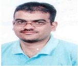
Kamlesh Guleria has completed his PhD from Guru Nanak Dev University, Amritsar, Punjab, India and Postdoctoral studies from Institute of Genetic Medicine, Newcastle University, Newcastle upon Tyne (UK). He also worked as Research Scientist at Sir Ganga Ram Hospital, New Delhi, India. Since 2009, he is working as Assistant Professor, at Department of Human Genetics Guru Nanak Dev University, Amritsar. He has published more than 20 papers in reputed journals and has been serving as an Editorial Board Member of repute and he has secured extramural research grants from various funding agencies in India
Abstract
Angiogenesis is an essential process in tumor growth, provides potential routes for tumor dissemination and metastasis. Vascular endothelial growth factor (VEGF) is a potent regulator of angiogenesis and is involved in the development and progression of solid tumors. Cancers use lymphangiogenesis and VEGF has been described as major mediator of breast cancer angiogenesis. The aim of the present study was to investigate the potential association of thirteen [ten promoter (-2578C/A, -2549I/D, -460T/C, -417T/C, -172C/A, -165C/T, -160C/T, -152G/A, -141A/C and -116G/A), two 5′-UTR (+405C/G, -7C/T) and one 3′-UTR (+936C/T)] VEGF polymorphisms with breast cancer risk in patients from Punjab, North-west India. In this study, DNA samples of 250 pathologically confirmed breast cancer patients and 250 ages and gender matched unrelated healthy individuals were screened for thirteen selected VEGF polymorphisms using direct PCR, PCR-RFLP and Sanger sequencing methods. Of 250 breast cancer patients, 7 were males and 243 were females. About 73.6% of patients developed breast cancer after 40 years of age. A significant association of VEGF -2578AA, -2549II, -460CC, +405GG, -152AA and -116AA genotypes with increased breast cancer risk were observed. VEGF -165CT and -141AC genotypes were associated with decreased risk for breast cancer. A strong linkage was observed between VEGF -2578C/A and -2549I/D, -2578C/A and -460T/C, -2549I/D and -460T/C and -165C/T and -141A/C polymorphisms. Haplotype analyses indicated that VEGF -2578A/-2549I/-460C/+405G/-7C/+936C and -417T/-172C/-165C/-160C/-152A/-141A/-116A combination was associated with increased risk while combination of -2578C/-2549D/-460T/+405G/-7C/+936C was associated with decreased risk for breast cancer. Analysis using TFSEARCH software revealed that VEGF -2578C/A, -417T/C, -172C/A and +405C/G polymorphisms altered the binding site of specific transcription factors. In this case-control study, VEGF -2578C/A, -2549I/D, -460T/C, +405C/G, -165C/T, -152G/A, -141A/C and -116G/A polymorphisms were associated with susceptibility to breast cancer in patients from Punjab, North-west India. VEGF is an important target in anti-cancer therapy, and findings about polymorphisms influencing VEGF-targeted therapies will help physicians to tailor therapy in individual manner.